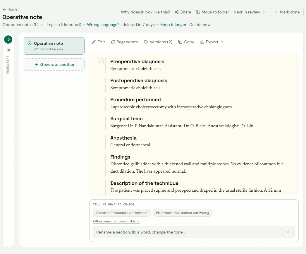
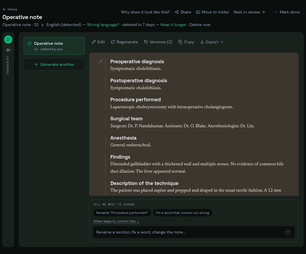
Preoperative diagnosis
Symptomatic cholelithiasis. (appears in this excerpt)
Postoperative diagnosis
Symptomatic cholelithiasis. (appears in this excerpt)
Procedure performed
Laparoscopic cholecystectomy with intraoperative cholangiogram. (appears in this excerpt)
Surgical team
Surgeon: Dr. P. Nandakumar. Assistant: Dr. O. Blake. Anesthesiologist: Dr. Lin. (appears in this excerpt)
Anesthesia
General endotracheal. (appears in this excerpt)
Findings
Distended gallbladder with a thickened wall and multiple stones. No evidence of common bile duct dilation. The liver appeared normal.
Description of the technique
The patient was placed supine and prepped and draped in the usual sterile fashion. A 12 mm umbilical port was placed by open technique and pneumoperitoneum established to 15 mmHg. Three additional 5 mm ports were placed under direct vision.
The gallbladder was retracted cephalad and the triangle of Calot dissected. The cystic duct and cystic artery were identified and the critical view of safety obtained. A cholangiogram through the cystic duct showed no filling defects. The cystic duct and artery were each clipped twice proximally and once distally and divided.
The gallbladder was dissected from the liver bed with electrocautery and removed in a retrieval bag through the umbilical port. Hemostasis was verified. The umbilical fascia was closed with 0 Vicryl and the skin with 4-0 Monocryl.
Specimens
Gallbladder to pathology.
Estimated blood loss and transfusions
20 mL. No transfusion.
Complications and incidents
None.
Counts
Correct times two.
Intraoperative studies
Cholangiogram: normal.
Immediate postoperative condition and plan
Extubated in the operating room, hemodynamically stable, to recovery.
Clear liquids tonight, advance as tolerated.
Oxycodone 5 mg every 6 hours as needed.
Discharge tomorrow if tolerating diet; follow-up in 2 weeks.